VCL offers a wide variety of animal models for different ocular pathologies:
- Age related Macular Degeneration
- Uveitis
- Glaucoma
- Diabetic retinopathy
VCL uses the eye as a model system to obtain insights into mechanisms and processes underlying many human diseases, i.e. pathological angiogenesis, fibrosis, inflammation and neurodegeneration.The models are listed below according to the different pathological processes that are involved.
VCL can help you in selecting the right models for testing of your compounds.
Angiogenesis and Vessel Leakage
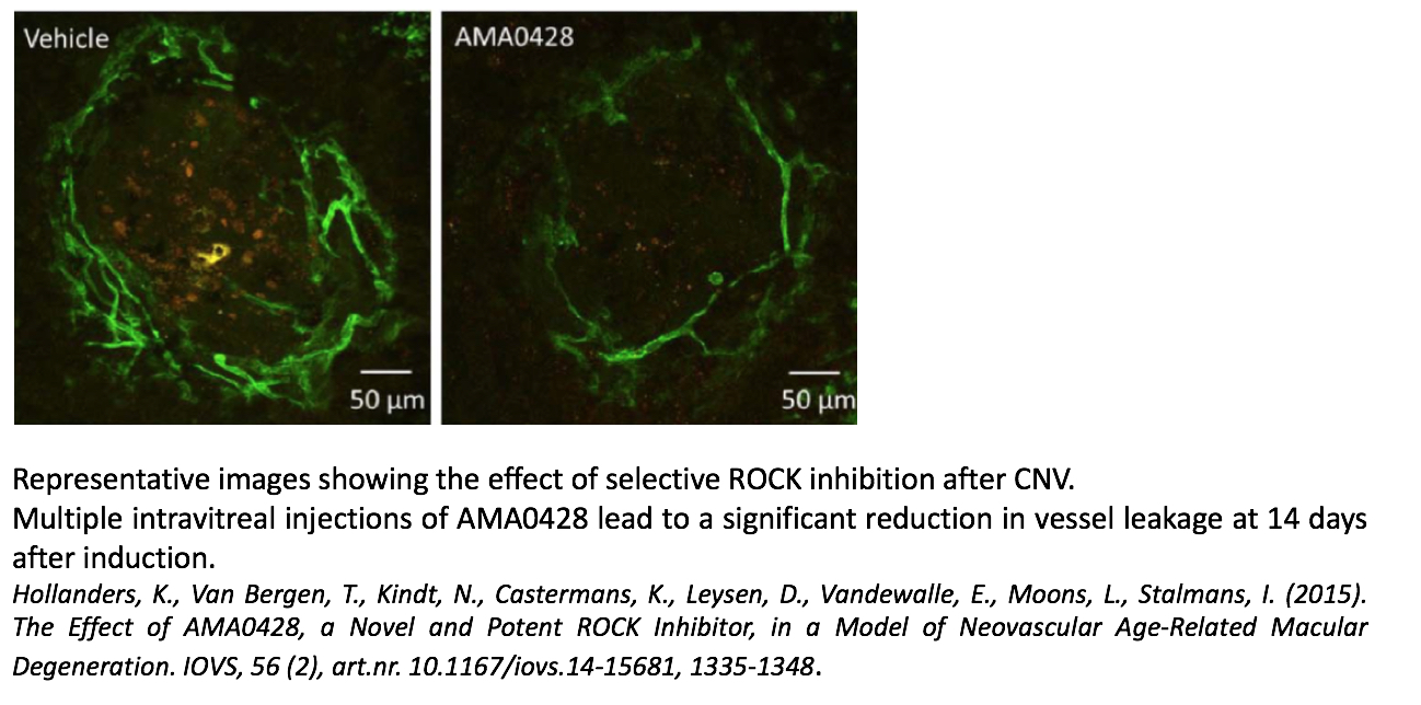
- Laser-induced choroidal neovascularization (AMD)
- Streptozotocin-induced diabetic retinopathy
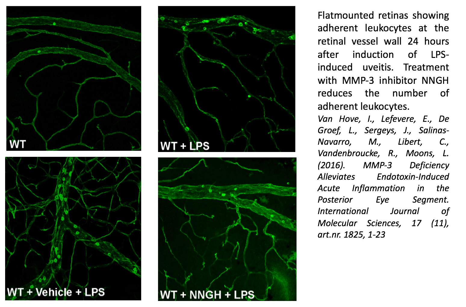
Inflammation
- Experimental autoimmune uveitis mouse model (EAU)
- Endotoxin-induced uveitis mouse model (EIU)
- Laser-induced choroidal neovascularization (AMD)
- Streptozotocin-induced diabetic retinopathy
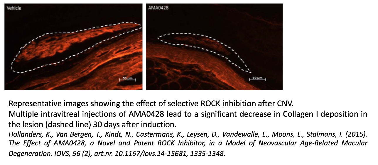
Fibrosis
- Fibrosis after ocular surgery
- Laser-induced choroidal neovascularization (AMD)
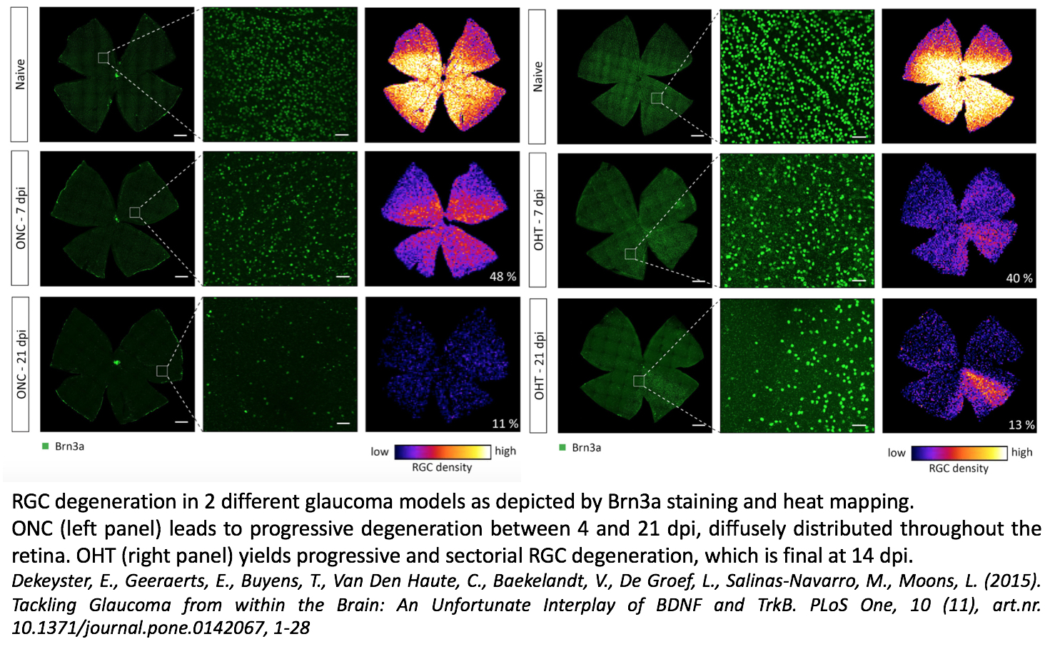
Neurodegeneration
- Microbead occlusion model
- Laser-induced ocular hypertension (glaucoma)
- Optic Nerve Crush (glaucoma)
- NMDA-induced excitotoxicity (glaucoma)
- Streptozotocin-induced diabetic retinopathy